Images of DDIT3


XL DDIT3 BAXL DDIT3 BADescriptionClinical DetailsImagesExpected PatternsLiteratureDownloadsRelated PublicationsRelated News


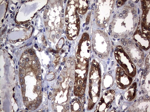
Immunohistochemistry (Formalin/PFA-fixed paraffin-embedded sections) - Anti-DDIT3 (phospho S30) antibody (ab63392)
Flow cytometric analysis of HepG2 cells with DDIT3 antibody at 1/50 dilution (fuchsia) compared with an unlabelled control (cells without incubation with primary antibody; yellow). Alexa Fluor 488-conjugated goat anti rabbit IgG was used as the secondary antibody.
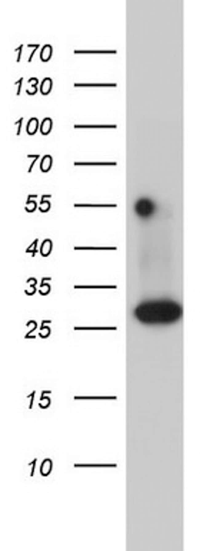
Western blot analysis of DDIT3 on different lysates. Proteins were transferred to a PVDF membrane and blocked with 5% BSA in PBS for 1 hour at room temperature. The primary antibody (ET1703-05, 1/500) was used in 5% BSA at room temperature for 2 hours. Goat Anti-Rabbit IgG - HRP Secondary Antibody (HA1001) at 1:200,000 dilution was used for 1 hour at room temperature.
Positive control:
Lane 1: LOVO cell lysate
Lane 2: PC-12 cell lysate
Predicted band size: 19 kDa
Observed band size: 25 kDa