Images of スペクトル幅

Mackie Quad Eq 4 Channel Graphic Equalizer And Real Time Spectrum Analyzer Ebay Spectrum Analyzer 4 Channel Spectrum

SiOWfa16: Science in Our World: Certainty and ControversyDoes the usage of microwave affect our health?
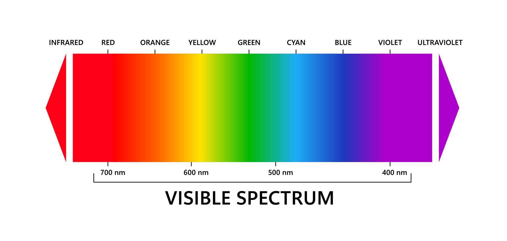
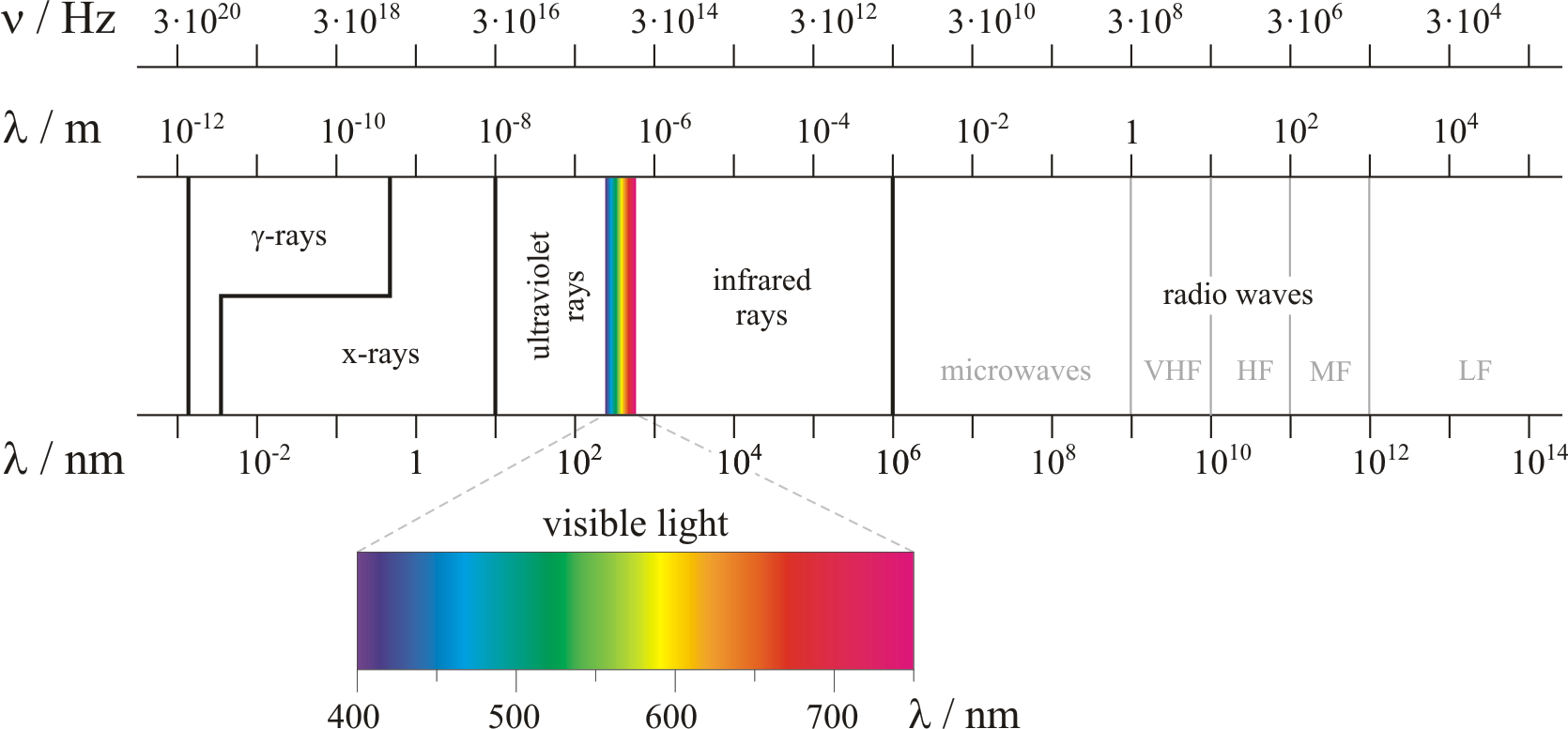
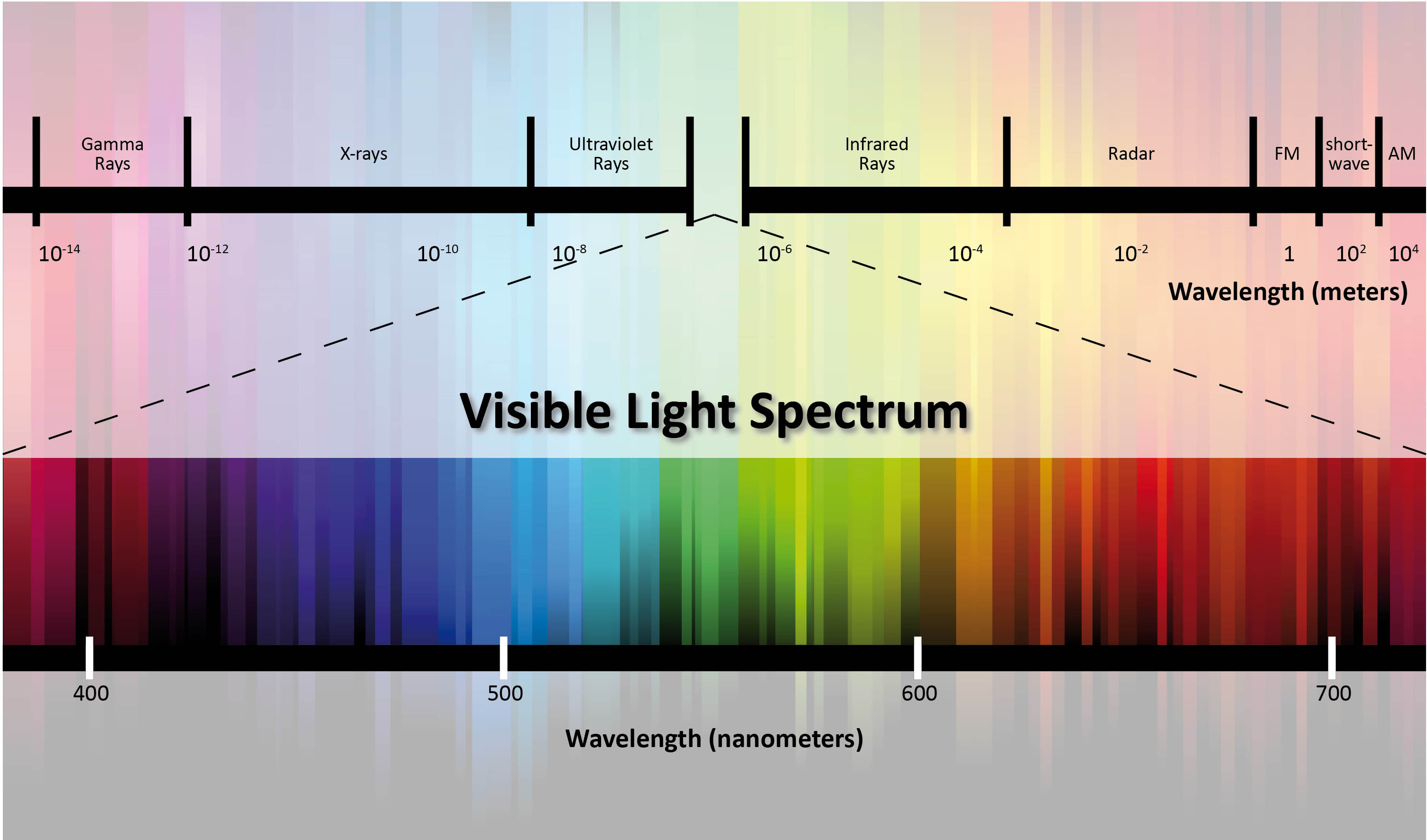
Visible light spectrum, infared and ultraviolet. Electromagnetic visible color spectrum for human eye. Vector gradient diagram with wavelength and colors. Educational illustration on white background
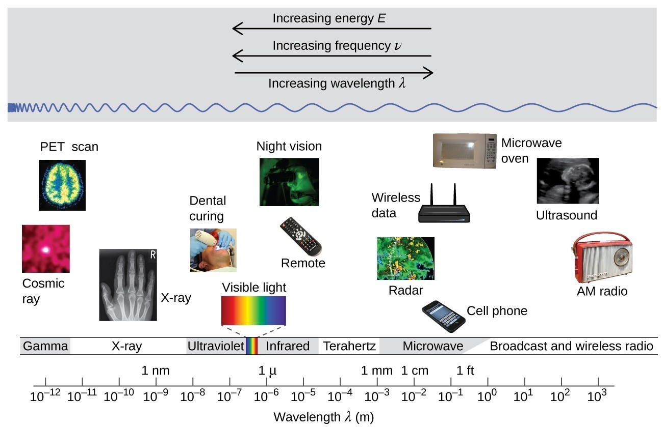
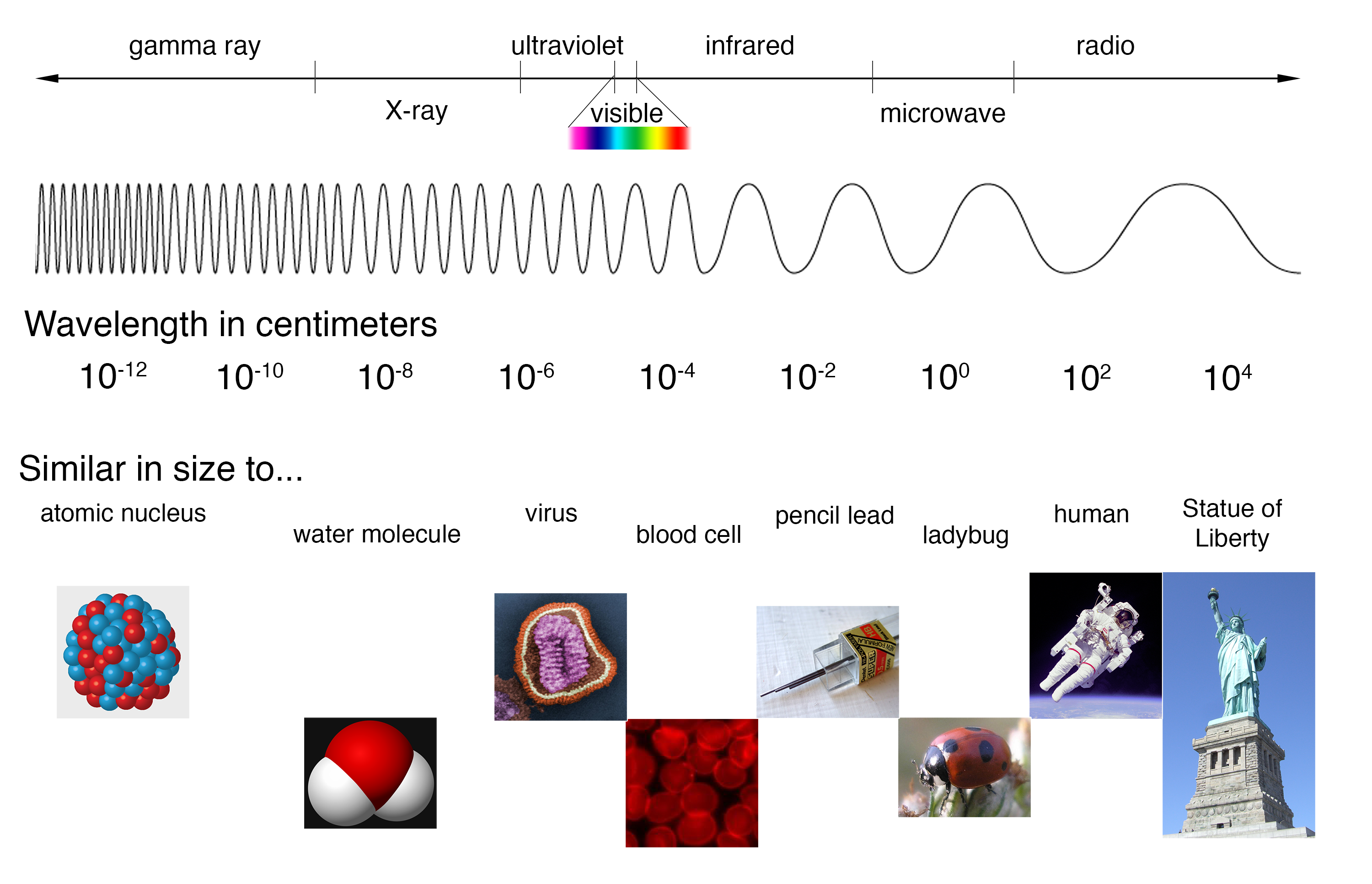
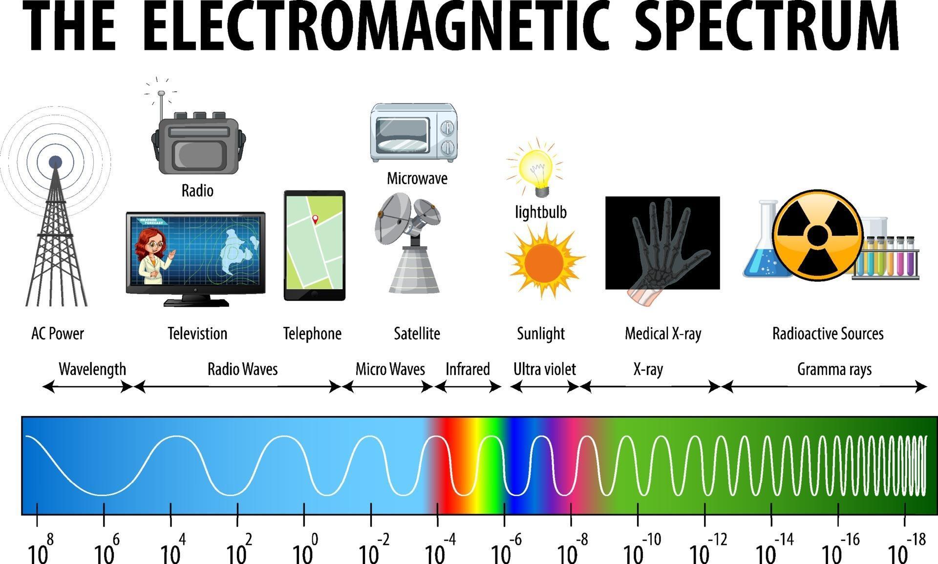
The figure includes a portion of the electromagnetic spectrum which extends from gamma radiation at the far left through x-ray, ultraviolet, visible, infrared, terahertz, and microwave to broadcast and wireless radio at the far right. At the top of the figure, inside a grey box, are three arrows. The first points left and is labeled, “Increasing energy E.” A second arrow is placed just below the first which also points left and is labeled, “Increasing frequency nu.” A third arrow is placed just below which points right and is labeled, “Increasing wavelength lambda.” Inside the grey box near the bottom is a blue sinusoidal wave pattern that moves horizontally through the box. At the far left end, the waves are short and tightly packed. They gradually lengthen moving left to right across the figure, resulting in significantly longer waves at the right end of the diagram. Beneath the grey box are a variety of photos aligned above the names of the radiation types and a numerical scale that is labeled, “Wavelength lambda ( m ).” This scale runs from 10 superscript negative 12 meters under gamma radiation increasing by powers of ten to a value of 10 superscript 3 meters at the far right under broadcast and wireless radio. X-ray appears around 10 superscript negative 10 meters, ultraviolet appears in the 10 superscript negative 8 to 10 superscript negative 7 range, visible light appears between 10 superscript negative 7 and 10 superscript negative 6, infrared appears in the 10 superscript negative 6 to 10 superscript negative 5 range, teraherz appears in the 10 superscript negative 4 to 10 superscript negative 3 range, microwave infrared appears in the 10 superscript negative 2 to 10 superscript negative 1 range, and broadcast and wireless radio extend from 10 to 10 superscript 3 meters. Labels above the scale are placed to indicate 1 n m at 10 superscript negative 9 meters, 1 micron at 10 superscript negative 6 meters, 1 millimeter at 10 superscript negative 3 meters, 1 centimeter at 10 superscript negative 2 meters, and 1 foot between 10 superscript negative 1 meter and 10 superscript 0 meters. A variety of images are placed beneath the grey box and above the scale in the figure to provide examples of related applications that use the electromagnetic radiation in the range of the scale beneath each image. The photos on the left above gamma radiation show cosmic rays and a multicolor PET scan image of a brain. A black and white x-ray image of a hand appears above x-rays. An image of a patient undergoing dental work, with a blue light being directed into the patient's mouth is labeled, “dental curing,” and is shown above ultraviolet radiation. Between the ultraviolet and infrared labels is a narrow band of red, orange, yellow, green, blue, indigo, and violet colors in narrow, vertical strips. From this narrow band, two dashed lines extend a short distance above to the left and right of an image of the visible spectrum. The image, which is labeled, “visible light,” is just a broader version of the narrow bands of color in the label area. Above infrared are images of a television remote and a black and green night vision image. At the left end of the microwave region, a satellite radar image is shown. Just right of this and still above the microwave region are images of a cell phone, a wireless router that is labeled, “wireless data,” and a microwave oven. Above broadcast and wireless radio are two images. The left most image is a black and white medical ultrasound image. A wireless AM radio is positioned at the far right in the image, also above broadcast and wireless radio.
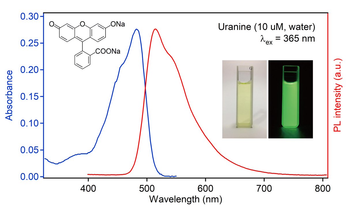
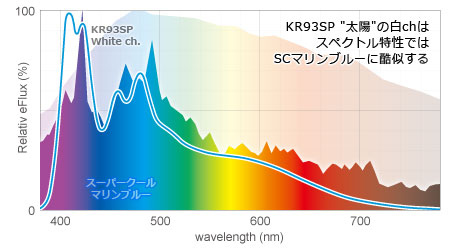
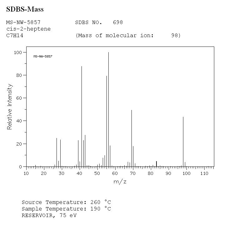
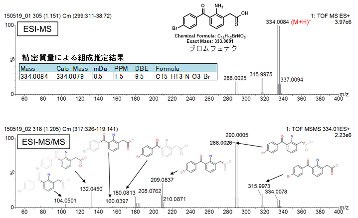
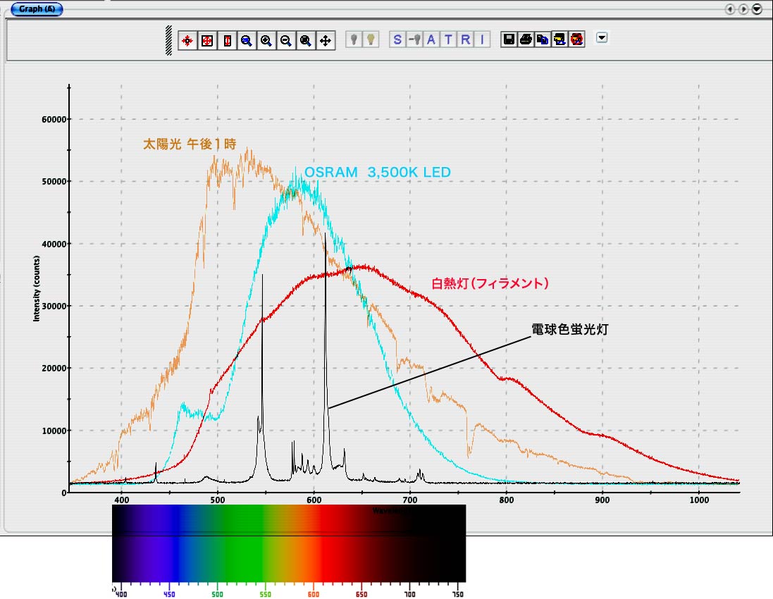
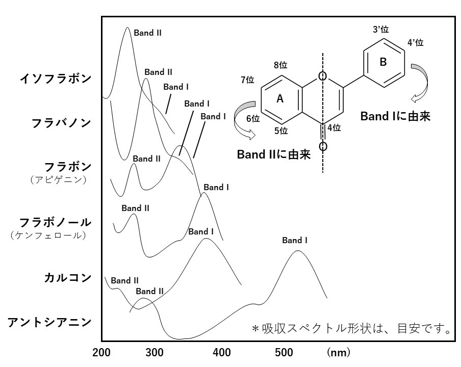
腫瘍判別用pH応答性シアニン系近赤外色素圧縮センシングの復元ダイナミックス解析図の書き方(作成中)J08 IV族およびIII-V族半導体の顕微フォトルミネセンス 東京大学大学院OITDA AM 01 (201x Ed. 1) 利得過渡パラメータに関する測定方法株式会社 サイアロン サイアロン蛍光体の特徴IR-VASETM - テクノ・シナジー当日配布資料光学工房 - 日本光学会SPARTANによる 紫外可視光吸収スペクトルの予測 - Wavefunction, Inc.紫外・可視スペクトル(UV-vis)高蛍光性含フッ素ポリイミド - 光吸収・蛍光特性のジアミン依存性 -PowerPoint プレゼンテーションUV-Vis スペクトル法S8.3 酸・塩基の硬さ・軟らかさ:HSAB 則 ブレンステッド酸塩基反応がTHz用材料とコンポーネントについてはこちらexpydoc.comYour ExpyDoc

![石目調 コレクションケース led 横ライト 幅50 奥行25 ミドルタイプ 薄型 コレクションボード ガラスケース キュリオケース 収納ケース ガラス 棚 ガラス棚 収納 キャビネット ガンプラ フィギュア オフィス [スペクトル]](https://thumbnail.image.rakuten.co.jp/@0_mall/cs-with/cabinet/item/spectral/y/spectral_y_50m_sts.jpg?_ex=300x300)


![アウトレット品 コレクションケース led 横 幅75 奥行25 ロータイプ 薄型 コレクションボード ガラスケース キュリオケース 収納ケース ディスプレイ ガンプラ フィギュア ウィスキー トミカ オフィス [スペクトル]](https://thumbnail.image.rakuten.co.jp/@0_mall/cs-with/cabinet/item/spectral/y/csw_spct75l_y_sowhwh.jpg?_ex=300x300)


![石目調 コレクションケース led 横ライト 幅75 奥行25 ロータイプ 薄型 コレクションボード ガラスケース キュリオケース 収納ケース 収納 ガンプラ フィギュア ウィスキー トミカ オフィス ガラス 棚 [スペクトル]](https://thumbnail.image.rakuten.co.jp/@0_mall/cs-with/cabinet/item/spectral/y/spectral_y_75l_sts.jpg?_ex=300x300)




![アウトレット品 コレクションケース led 横ライト 幅50 奥行25 ミドルタイプ 薄型 コレクションボード ガラスケース キュリオケース 収納ケース ガラス棚 収納 ディスプレイ ガンプラ フィギュア オフィス [スペクトル]](https://thumbnail.image.rakuten.co.jp/@0_mall/cs-with/cabinet/item/spectral/y/spectral_y_50m_olt_s.jpg?_ex=300x300)



![コレクションケース led 横ライト 幅75 奥行25 ロータイプ 薄型 コレクションボード ガラスケース キュリオケース 収納ケース 収納 ガンプラ フィギュア ウィスキー トミカ オフィス ガラス 棚 [スペクトル]](https://thumbnail.image.rakuten.co.jp/@0_mall/cs-with/cabinet/item/spectral/y/spectral_y_75l_s.jpg?_ex=300x300)


![石目調 コレクションケース led 横ライト 幅50 奥行25 ハイタイプ 薄型 コレクションボード ガラスケース 飾り棚 キュリオケース 収納ケース ガラス ガラス棚 キラキラ収納 ガンプラ フィギュア オフィス[スペクトル]](https://thumbnail.image.rakuten.co.jp/@0_mall/cs-with/cabinet/item/spectral/y/cs_spctl_y_50hst_s.jpg?_ex=300x300)

